Diseases We Treat
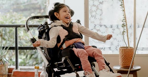
Cerebral Palsy
Cerebral palsy, a neurological condition, happens either due to brain damage during fetal development or because of another developmental disability that impacts the brain’s growth.
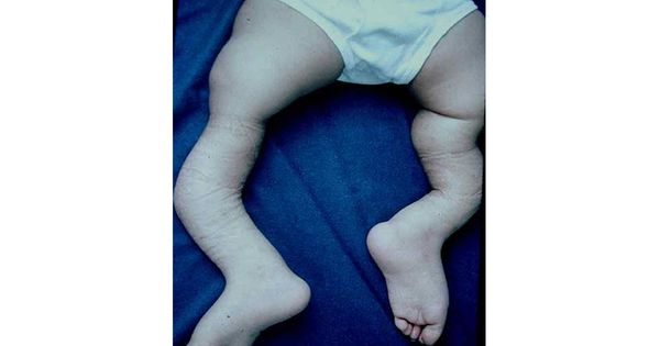
Osteogenesis Imperfecta
Osteogenesis Imperfecta (OI), or Brittle Bone Disease, is a condition where the person has fragile bones that break easily with no evident reason.
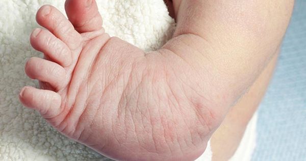
Clubfoot - CTEV
It is the most common congenital deformity of the foot that affects a child’s muscles, bones, tendons, and blood vessels.
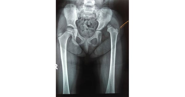
DDH (Developmental dysplasia of the hip)
DDH is when the baby's hip joints do not form well. In this condition, the ball part of the hip joint is partly or completely out of the socket.
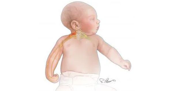
Erb's Palsy
Erb’s palsy refers to a muscle weakness in the arms or the shoulder, occurring as the after-effect of an injury to the brachial plexus at birth.
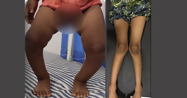
Genu Varum & Genu Valgum
Genu varum or Bow legs is a condition where a child’s legs curve outwards at the knees, creating a space between the knees and lower legs. The child’s legs appear like a bow when standing with feet and ankles together.
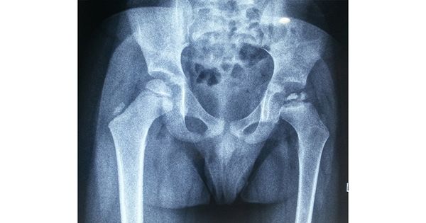
Perthes Disease
Perthes Disease, or Legg-Calve Perthes disease, is a condition that usually affects children of the age group 4-8 years old but can extend to two to 12 years old in some cases.
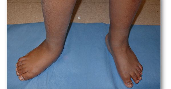
Tarsal Coalition (Rigid Flatfoot)
It is a condition where an abnormal connection is seen between the bones of the foot. Typically, it occurs before birth, with symptoms seen in late childhood or during teenage years.
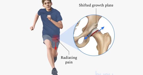
Slipped Capital Femoral Epiphysis - SCFE
Slipped Capital Femoral Epiphysis - SCFE
Hip Preservation Surgeries
Hip Preservation Surgeries
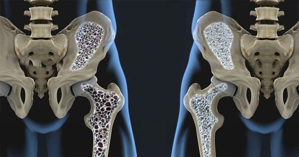
Metabolic Disorders like Rickets
Metabolic Disorders like Rickets
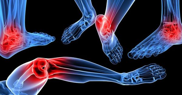
Foot and Ankle Disorders (Flatfeet, Tarsal Coalition)
Foot and Ankle Disorders as Flatfeet, Tarsal Coalition
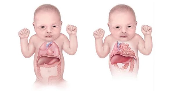
Congenital Anomalies
Congenital Anomalies